Basic Hair Structure: Hair Follicle and Hair Shaft Function Explained
By Paul Taylor

Hair structure and hair growth are surprisingly complex.
And perhaps that's partly why the exact mechanism causing hair loss has been a mystery for so long.
To help understand the hair loss process, it makes sense then to learn about the structure of hair and how it grows.
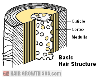
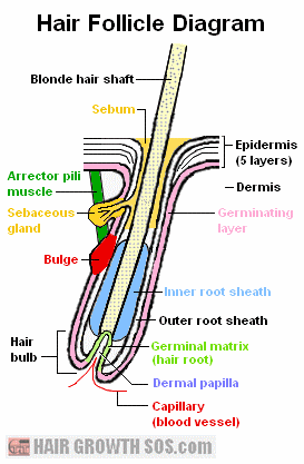
As you read, please refer the words in bold to the Basic Hair Structure and Hair Follicle diagrams below. Some parts of the Hair Follicle Diagram are also color-coded to highlight their importance.
Since hair growth is basically just a collection of dead cells being pushed along a
pouch (follicle) in the skin, it’s important to learn about
skin structure and function too.
Skin Structure and function
Skin is the largest organ in the body. It has various functions including:
- Temperature regulation (sweat glands to
cool down; goosebumps to keep warm).
- Excretion (the skin is sometimes
referred to as the "third kidney").
- Protection (against sun, rain, bugs, infection, etc).
Skin has two main parts - the epidermis and dermis.
The epidermis has five layers. The uppermost layer forms the surface of the skin and is made from dead cells which are continuously being shed and replaced from below. Excessive shedding can, of course, produce dandruff.
The deepest layer of the epidermis is the germinating layer. This region is constantly growing and dividing into new cells, pushing the old cells up towards the surface of the skin.
Below the epidermis is the dermis.
The dermis is the thickest part of the skin and contains blood vessels
to supply the nutrients needed for skin cells to grow.
Basic Hair Structure and function
Before hair growth can begin, a hair follicle must first be created:
The germinating layer of the epidermis starts growing down into the dermis, and forms the outside of each hair follicle.
The dermis then grows upwards into the base of the follicle to form the dermal papilla. This allows capillaries (blood vessels) to enter the papilla and provide nutrients for the hair shaft to grow.
The bottom part of the follicle enlarges into an
area of actively growing cells. This is called the hair bulb.
At the base of the hair bulb, the germinating layer merges into the outer root sheath (which forms the inner wall of the follicle).
The outer root sheath then forms the germinal matrix (hair root) which surrounds the dermal papilla.
The germinal matrix grows the inner root sheath (this is the white bit at the end of a hair if it's pulled out).
The germinal matrix also contains stem cells - these grow the hair shaft through constant cell division which continuously push older cells upwards.
Hair shaft cells are similar at first. But as they move up through the follicle, they begin to change shape, and a protein called keratin develops inside the cells.
Three different types of hair cell then form (see hair shaft structure diagram below). By the time these cells are a third of the way up the follicle, they have died and fully hardened (keratinized).
A sebaceous gland lies within each follicle. This produces an oily substance called sebum from a duct which opens up into the hair follicle about halfway down from the skin surface.
The follicle also has a bulge directly below the sebaceous gland in the outer root sheath at the attachment point of the arrector pili muscle*.
The bulge produces stem cells which regenerate the follicle during the next hair growth cycle.
* When the arrector pili
muscles contract, they make your hair stand on end. This is what causes goosebumps.
Hair Shaft Structure


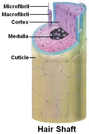
In the basic hair structure diagram above, you can see that the hair shaft has three layers: the cuticle (outer layer), cortex (middle layer) and medulla (inner layer).
The medulla is a honeycomb keratin structure with air spaces inside.
The cortex gives flexibility and tensile (stretching) strength to hair and contains melanin granules, which give hair its color (blond in the diagram above).
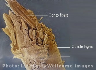
The cortex is made from tiny fibers of keratin running parallel to each other along the length of the hair shaft (as shown in the photo of the split hair end above).
The cuticle is made from 6 to 11 layers of overlapping semi-transparent keratin scales (which make the hair waterproof and allow it to be stretched).
Someone who has thick,
course hair will have more overlapping layers of cuticles than someone
with fine hair. You can just see the cuticle layers in the photo above. And in the diagram, you can count 6 cuticle layers.
Take a Closer Look
Beneath the skin of keratin
As already stated, hair cells are mostly made from keratin (up to 95%). And, like all proteins, keratin is made from amino acids.
Sixteen amino acids form keratin, with cysteine having the highest concentration. And cysteine is one of just two amino acids that have a high sulfur content (the other being methionine).
The significance of all this is that the mineral sulfur is known to be beneficial for hair growth. Clearly then, sulfur forms a very important component of hair structure.
Different type of hair: Same type of hair structure
There are three types of hair - lanugo, vellus and terminal. All of them share a similar basic hair structure, but each plays a different role in the body:
Lanugo hair starts growing when you’re still in the womb, but falls out either just before or just after birth. (So, in other words, you might experience hair loss before you’ve even been born!)
After your lanugo hair falls out, it gets replaced by vellus hair - this is the tiny, near invisible hair that covers your body.
However, a lot of vellus hair turns into terminal hair (which includes eyebrows, eyelashes and scalp hair). And male hormones (androgens) also stimulate vellus hair to "mature" into facial hair, body hair and pubic hair.
Paradoxically, the very same androgens associated with hair growth, have also been associated with hair loss (androgenetic alopecia). These include testosterone and dihydrotestosterone (DHT). So, it’s important to take a look at where within the hair follicle these androgens act.
Which hair structures are connected to hair loss?
People with hair loss tend to have higher levels of 5-alpha reductase and androgen receptor sites in the hair follicles within the hair loss region of the scalp (1).
5-alpha reductase is an enzyme that converts testosterone into DHT. And androgen receptor sites are essentially attachment points for androgens (especially DHT).
In hair follicles, 5-alpha reductase is found in the sebaceous glands, dermal papilla, inner root sheath and outer root sheath (2). And androgen receptor sites are found in dermal papilla cells and sebaceous glands (3).
As you can see, these two important components of the hair loss process (5-alpha reductase and androgen receptor sites) exist within various structures of the hair follicle.
And this, of course, only adds to the complexity this puzzling type of hair loss presents.
However, it's a puzzle that, I believe, has now been solved. Read the skull expansion section to learn more.
You now know all about the various structures which form the hair follicle and hair shaft. On the next page, learn how all these different parts of the hair follicle interact to create the hair on your head over and over again throughout life.
This is page 1 of 3.
Read next
page? Hair growth cycle.
|
Like this page? |
|


